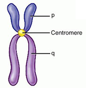
In the nucleus of each cell, the DNA molecule is packaged into thread-like structures called chromosomes. Each chromosome is made up of DNA tightly coiled many times around proteins called histones that support its structure.

Chromosomes are not visible in the cell’s nucleus—not even under a microscope—when the cell is not dividing. However, the DNA that makes up chromosomes becomes more tightly packed during cell division and is then visible under a microscope. Most of what researchers know about chromosomes was learned by observing chromosomes during cell division.

Image is a chromosome with the centromere labeled

Source: Chromosome, ingmarz, All Strangled Up: A Blog about DNA and DNA Research

Each chromosome has a constriction point called the centromere, which divides the chromosome into two sections, or “arms.” The short arm of the chromosome is labeled the “p arm.” The long arm of the chromosome is labeled the “q arm.” The location of the centromere on each chromosome gives the chromosome its characteristic shape, and can be used to help describe the location of specific genes.

To "read" a set of human chromosomes, scientists first use three key features to identify their similarities and differences:

  1. Size. This is the easiest way to tell two different chromosomes apart.
  2. Banding pattern. The size and location of Giemsa bands on chromosomes make each chromosome pair unique.
  3. Centromere position. Centromeres are regions in chromosomes that appear as a constriction. They have a special role in the separation of chromosomes into daughter cells during mitosis cell division (mitosis and meiosis).

Using these key features, scientists match up the 23 pairs—one set from the mother and one set from the father.

Source: How to read a chromosome, University of Utah: Learn Genetics

Chromosomes can be studied by creating a karyotype. In order to do this, cells from the organism are grown in a laboratory. After the cells have reproduced a few times, they are treated with a chemical that stops cell division in the middle phase. During this middle phase, the chromosomes are at the best length for identification. The cells are treated further, stained, and then placed on a glass slide. The chromosomes are observed under the microscope where they are counted and photographed. The photograph is then enlarged, and the chromosomes are individually cut out. The chromosomes are identified and arranged in homologous pairs. Homologous chromosomes are identical, or matching, chromosomes. One chromosome in a homologous chromosome pair comes from the mother, the other from the father. The arrangement of homologous chromosome pairs is called a karyotype. Humans have 46 chromosomes, 23 pairs. A human karyotype would show 23 pairs of homologous chromosomes, lined up from largest to smallest. 44 chromosomes, 22 pairs, are autosomes, body chromosomes. The other pair is called the sex chromosomes because they determine the gender or sex of an individual. The presence of a Y chromosome means the individual is a male.

Image Adapted From: Normal Male Karyotype, Biology Corner